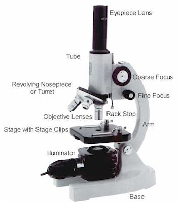
The stage is the rectangular platform upon which the microscope slide is placed.(This passage was adapted from Microbiology: A Laboratory Manual,5th edition, Cappuccino, J.S. The base is the support upon which the instrument rests. Parts of the Compound Microscope 1. Lock the cabinet with the combination padlock, keyhole facing outwards. Pull the dust cover all the way over the base.
It also carriers the microscopic illuminators.Guide to microscopes, including types of microscopes, parts of the microscope, general use and. Base It acts as microscopes support. Head This is also known as the body, it carries the optical parts in the upper part of the microscope. There are three structural parts of the microscope i.e.
The former use visible light orultraviolet rays to illuminate specimens. Microscopes are designated as either lightmicroscopes or electron microscopes. Over the years, microscopes have evolved from the simple,single-lens instrument of Leeuwenhoek, with a magnification of 300,to the present-day electron microscopes capable of magnificationsgreater than 250,000. In 1673, with the aid of a crude microscopeconsisting of a biconcave lens enclosed in two metal plates,Leeuwenhoek introduced the world to the existence of microbial formsof life. Here youll find all the.3.To learn the correct use of the microscope for observation andmeasurement of microorganisms.Microbiology, the branch of science that has so vastly extendedand expanded our knowledge of the living world, owes its existence toAntony van Leeuwenhoek. But if you buy a quality one and maintain it well, it will last a lifetime.
The specimen is illuminated by a beam oftungsten light focused on it by a sub-stage lens called a condenser,and the result is that the specimen appears dark against a brightbackground. Electron microscopes use elec-tron beams instead of lightrays, and magnets instead of lenses to observe submicro-scopicparticles.Essential Features of Various MicroscopesThis instrument contains two lens systems for magnifyingspecimens: the ocular lens in the eyepiece and the objective lenslocated in the nose-piece. Fluorescentmicro-scopes use ultraviolet radiations whose wavelengths are shorterthan those of visible light and are not directly perceptible to thehuman eye.
The special optics convert the difference betweentransmitted light and refracted rays, resulting in a significantvari-ation in the intensity of light and thereby producing adiscernible image of the struc-ture under study. As light is transmitted througha specimen with a refractive index different from that of thesurrounding medium, a portion of the light is refracted (bent) due toslight varia-tions in density and thickness of the cellularcomponents. Its optics include special objectives and acondenser that make visible cellular components that differ onlyslightly in their refractive indexes. Living specimens may beobserved more readily with darkfield than with brightfieldmicroscopy.Observation of microorganisms in an unstained state is possiblewith this microscope. The con-denser directs the light obliquely so that thelight is deflected or scattered from the spec-imen, which thenappears bright against a dark background. Therefore, most brightfieldobservations are performed on nonviable, stained preparations.This is similar to the ordinary light microscope however, thecondenser system is modified so that the specimen is not illuminateddirectly.
Ultraviolet radiations are absorbed by the fluorescentlabel and the energy is re-emitted in the form of a differentwavelength in the visible light range. The ocular lensis fitted with a filter that permits the longer ultravioletwavelengths to pass, while the shorter wavelengths are blocked oreliminated. The source ofillumination is an ultraviolet (UV) light obtained from ahigh-pressure mercury lamp or hydrogen quartz lamp.
These components aresealed in a tube in which a complete vacuum is established.Transmission electron microscopes require speci-mens that are thinlyprepared, fixed, and dehydrated for the electron beam to pass freelythrough them. In theelectron microscope, the specimen is illu-minated by a beam ofelectrons rather than light, and the focusing is carried out byelec-tromagnets instead of a set of optics. This permits visualization ofsubmicroscopic cel-lular particles as well as viral agents. Antibodies are conjugatedwith a fluorescent dye that becomes excited in the presence ofultraviolet light, and the fluorescent portion of the dye becomesvisible against a black background.This instrument provides a revolutionary method of microscopy,with magnifications up to one million. This microscope is used primarily for thedetection of antigen-antibody reactions.
Therefore, only the compound brightfield microscope willbe discussed in depth and used to examine specimens.1.Theoretical principles of brightfield microscopy.2.Component parts of the compound micro-scope.3.Use and care of the compound microscope.4.Practical use of the compound microscope for visualization ofcellular morphology from stained slide preparations.Microbiology is a science that studies living organisms that aretoo small to be seen with the naked eye. Although you should befamiliar with the basic principles of microscopy, you probably havenot been exposed to this diverse array of complex and expensiveequipment. Scanning electronmicroscopes are used for visualizing surface characteristics ratherthan intracellular structures A narrow beam of electrons scans backand forth, producing a three-dimensional image as the electrons arereflected off the specimen's surface.While scientists have a variety of optical instruments with whichto perform routine laboratory procedures and sophisticated research,the compound brightfield micro-scope is the "workhorse" and iscommonly found in all biological laboratories.
The flatside of the mirror is used for artificial light, and the concave sidefor sunlight.This component is found directly under the stage and contains twosets of lenses that collect and concentrate light passing upward fromthe light source into the lens sys-tems. Others are provided with a mirror oneside flat and the other concave.An external light source, such as a lamp, is placed in front ofthe mirror to direct the light upward into the lens system. Less sophisticated micro-scopes have clips on the fixedstage, and the slide must be positioned manually over the centralopening.The light source is positioned in the base of the instrument.Some microscopes are equipped with a built-in light source topro-vide direct illumination. Inaddition to the fixed stage, most microscopes have a mechanical stagethat can be moved vertically or horizontally by means of adjustmentcontrols. This platform provides a surface for theplacement of a slide with its specimen over the central opening. Althoughthere are many types and variations, they all fundamentally consistof a two-lens system, a variable but controllable light source, andmechanical adjustable parts for determining focal length between thelenses and specimen.A fixed platform with an opening in the center allows for thepassage of light from an illu-minating source below to the lenssystem above the stage.

When a lens cannotdiscriminate, that is, when the two objects appear as one, it haslost resolu-tion. Bydefinition, resolving power is the ability of a lens to show twoadjacent objects as discrete entities. When these are combined with themagnification of the ocular lens, the total or overall linearmagnification of the specimen is obtained.Although magnification is important, you must be aware thatunlimited enlargement is not possible by merely increasing themagnifying power of the lenses or by using additional lenses, becauselenses are limited by a property called resolving power. The objective lens is nearer thespecimen and magnifies it, producing the real image that is projectedup into the focal plane and then magnified by the ocular lens toproduce the final image.The most commonly used microscopes are equipped with a revolvingnosepiece containing four objective lenses possessing differentdegrees of magnification. These lensesare separated by the body tube. The body tube may be raised or lowered withthe aid of coarse-adjustment and fine-adjustment knobs that arelocated above or below the stage, depending on the type and make ofthe instrument.To use the microscope efficiently and with minimal frustration,you should understand the basic principles of microscopy:magnification, resolution, numerical aperture, illumination, andfocusing.Enlargement or magnification of a specimen is the function of atwo-lens system the ocular lens is found in the eyepiece, and theobjective lens is situated in a revolving nose-piece.


 0 kommentar(er)
